Amyloid and Tau PET imaging advance Alzheimer's disease diagnosis
Molecular imaging allows clinicians to visualize amyloid-β plaques and tau tangles in living patients, shifting Alzheimer's diagnosis toward a biological model.
- Headline Dispatch: Amyloid and Tau PET imaging advance Alzheimer's disease diagnosis
- Core Takeaway: Molecular imaging allows clinicians to visualize amyloid-β plaques and tau tangles in living patients, shifting Alzheimer's diagnosis toward a biological model.
- Source Synthesis: Continuous live monitoring aggregated across major news wires and independent bureaus.
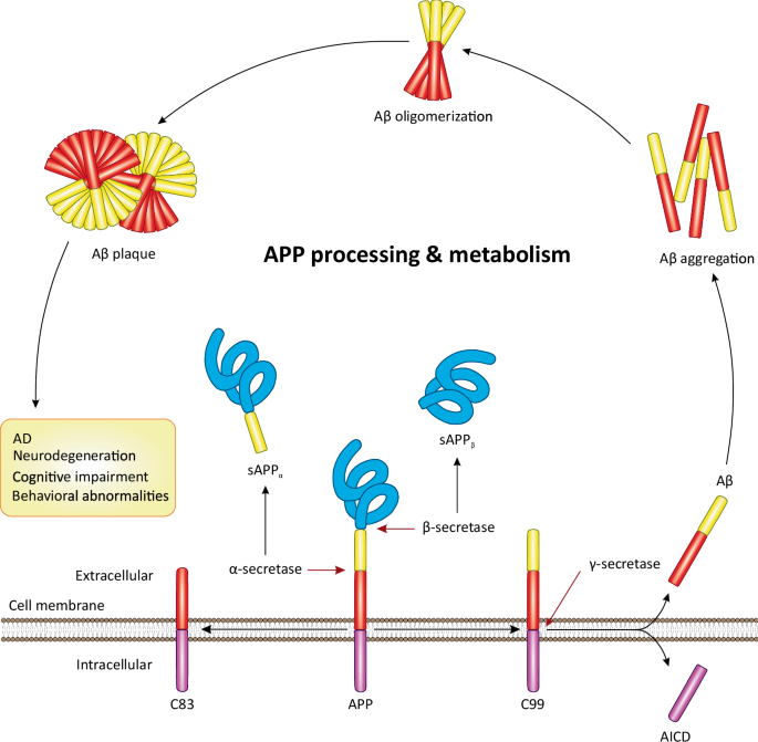
Amyloid and Tau PET imaging advance Alzheimer's disease diagnosis
The diagnostic approach to Alzheimer’s disease (AD) is shifting from a reliance on clinical symptoms to a biologically based model using molecular imaging. Positron emission tomography (PET) now allows clinicians to visualize the hallmark pathological proteins of the disease — amyloid-β (Aβ) plaques and tau tangles — while patients are still living.
Alzheimer’s is the most prevalent neurodegenerative disease and the leading cause of dementia worldwide. Traditionally, the disease was confirmed only postmortem. However, clinical diagnoses made before death have historically been unreliable, with some studies reporting misdiagnosis rates of 25%–30% in dementia patients.
The Role of Amyloid PET
Amyloid PET imaging targets the extracellular Aβ plaques that characterize AD. While PiB is widely used in research, its requirement for an on-site cyclotron has limited its scalability. This led to the development of 18F-labeled tracers, which have longer half-lives and broader clinical availability. Three such tracers, [18F]florbetapir, [18F]florbetaben, and [18F]flutemetamol, have been approved by the Food and Drug Administration (FDA) and the European Medicines Agency (EMA).
The clinical importance of amyloid PET has increased with the arrival of monoclonal antiamyloid antibodies in the early 2020s. Therapies require biomarker-verified amyloid positivity before treatment can begin.
Tau PET and Disease Staging
While amyloid plaques often appear early, tau PET provides a more direct correlate to clinical severity and disease stage. Tau pathology involves the intracellular accumulation of hyperphosphorylated tau into neurofibrillary tangles (NFTs). These tangles typically originate in the transentorhinal region of the medial temporal lobe, the dorsal raphe nucleus, and the locus coeruleus before spreading to limbic and isocortical regions.
First-generation tau tracers, such as [18F]flortaucipir, allow for the monitoring of tau spread. However, some first-generation tracers struggle to detect primary tauopathies like progressive supranuclear palsy (PSP) or corticobasal degeneration (CBD) and may suffer from off-target binding to monoamine oxidase B (MAO-B) in the basal ganglia. Second-generation tracers, including [18F]MK6240 and [18F]PM-PBB3, have been developed to improve binding affinity and selectivity.
Integrated Diagnostics and Metabolic Imaging
Beyond amyloid and tau, 18F-FDG PET is used to visualize neuronal dysfunction by identifying regional patterns of hypometabolism. In AD patients, hypometabolism often appears in the precuneus, posterior cingulate, and temporoparietal cortices. This specific pattern can create a visual effect known as the earmuff
or headphone
sign.
Clinicians often integrate these PET modalities with other biomarkers:
- Fluid Biomarkers: Cerebrospinal fluid (CSF) ratios of Aβ42/Aβ40 and plasma p-tau (such as p-tau217) offer early detection but lack the spatial resolution of PET.
- Structural Imaging: T1-weighted MRI is used to detect regional atrophy in the medial temporal lobe.
- Quantitative Scales: The Centiloid scale provides a standardized linear transformation (from 0 to 100) to compare amyloid burden across different scanners and tracers.
Access and Risks
The adoption of these tools has been influenced by reimbursement policies. CMS expanded coverage for amyloid PET in 2023 and began reimbursing tau PET scans between 2024 and 2025.
Looking forward, the integration of AI-based quantitative tools and new tracers for synaptic density (such as [11C]UCB-J) may further refine the ability to diagnose AD in its preclinical stages and personalize treatment durations.
How significant is this development?
Participate in our community survey on the trajectory and real-world impact of this news.