Salk Institute identifies iron accumulation pathway linked to neurodegeneration
Scientists have discovered a cellular stress pathway where gradual iron buildup reduces neuron resilience, offering a new target for treating neurodegenerative disorders.
Salk Institute identifies iron accumulation pathway linked to neurodegeneration
Scientists at the Salk Institute have identified a chronic stress pathway in cells that reduces the resilience of neurons over time, leaving them more susceptible to neurodegenerative diseases. The process, which the researchers named chronoferroptosis
, explains how the gradual accumulation of iron in the brain weakens cellular defenses and increases vulnerability to stressors.
The findings were published in the journal Cell Death Discovery on June 18, 2026. The research suggests that iron accumulation in brain cells is a key target for those attempting to predict, prevent, and treat neurodegenerative disorders.
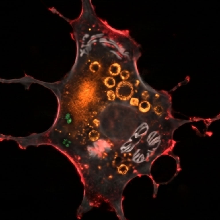
Iron is an essential mineral used for hormone production, energy production, the immune system, and the development of red blood cells. It is commonly found in seafood, lean meats, starchy cereals, and dark leafy greens. According to co-corresponding author Nawab John Dar, PhD, a postdoctoral researcher in the lab of Pam Maher, the problem is not the iron itself, but rather the accumulation of the mineral over time.
The progressive model of chronoferroptosis
While previous experiments typically examined iron exposure over 24- to 48-hour spans, the Salk team developed the first progressive model of iron accumulation using a human-derived nerve cell line. This allowed the researchers to compare acute exposure, lasting between six and eight hours, against chronic exposure lasting nine days.
The study revealed a stark difference between the two groups. In neurons exposed to iron acutely, there was very little biochemical difference before and after exposure. These cells were able to handle further stress.
In contrast, neurons subjected to chronic exposure exhibited significant changes, including:
- Elevated lipid peroxidation.
- The accumulation of harmful chemicals and the depletion of helpful ones.
- Upregulation of some cellular processes and downregulation of others.
These chronically exposed neurons remained viable but became hypersensitive to oxidative injury and were unable to handle additional stress. The researchers noted that this state does not necessarily end in immediate cell death but instead acts as a cellular stress pathway.
Mechanisms and cellular resilience
The discovery adds a temporal dimension to ferroptosis. Previously, ferroptosis was viewed strictly as an iron-dependent cell death pathway driven by lipid peroxidation. Senior and co-corresponding author Pam Maher, PhD, a research professor at Salk, compared this process to the way cooking oil or a nut goes bad through peroxidation.
The Salk team suspects that the buildup occurs because the iron export machinery in the cells fails. Under this theory, iron enters the neurons normally but cannot be removed after it has been used. The researchers are still investigating why this failure does not impact neurons for a significant period of time.
"Our study reveals that cells lose resilience when iron hits a certain level, making neurons more susceptible to stressors that damage or even kill them."
Pam Maher, PhD, research professor at Salk, via salk.edu
This loss of resilience is particularly relevant to the tens of millions of people worldwide affected by neurodegenerative diseases. In the United States, the Parkinson’s Foundation and Alzheimer’s Disease Association report roughly 1 million people with Parkinson’s and 7 million with Alzheimer’s.
Potential for future therapy
The research indicates that the fate of these cells is not sealed by the specific amount of iron present, but by the duration of time they spend under stress. This suggests a window for intervention if scientists can detect when the brain begins entering this vulnerable state.
While not the primary focus of the published paper, Maher stated that her lab has developed several compounds to inhibit this pathway. Such interventions could potentially address iron imbalances and boost neuron resilience to stave off neurodegeneration during aging.
The paper was coauthored by David Soriano-Castell of Salk and received support from the National Institutes of Health via grants R01AG067331 and R01AG069206.